Category: Practicals

  • RBC Count

    Aim: To find out the number of red blood cells in one cubic millimetre of blood.

    Principles: The number of RBC in a known volume of diluted blood is counted and the number of
    cells in one cm of undiluted blood are calculated from this.

    Apparatus Required: Hemocytometer, RBC diluting fluid, compound microscope, sterile lancet,
    watch glass, cotton, rectified spirit.

    Uses of R.B.C. pipette:

    • WBC count in leukemias
    • Platelet counting

    Uses of the bead in the bulb:

    • For proper mixing
    • To know whether the pipette is dry
    • To identify the pipette
    • R.B.C diluting fluid: Hayem’s fluid is the commonly used diluting fluid.

    Composition:

    • Sodium chloride 0.5 Gm
    • Sodium sulphate 2.5 Gm
    • Mercuric perchloride 0.25 Gm
    • Distilled water 100 ml sodium chloride and sodium sulphate together keep the isotonicity of fluid. Sodium sulphate also prevents the clumping of red cells. Mercuric perchloride fixes the cells and acts as a preservative.
    • Other diluting fluids:
      • Gower’s fluid
      • Toison’s fluid
      • Formol Citrate solution

    Procedure: Clean and dry the counting chamber and put on the special coverslip provided. Focus on the high power objective and identify the RBC counting area. Clean the RBC pipette first with distilled water, then with absolute alcohol and finally with ether and keep it dry. Take a small quantity of diluting fluid in a watch glass and keep it aside. Clean the fingertip using rectified spirit and make a deep prick with a sterile lancet, so that blood comes out freely without squeezing. Wipe off the first drop which may contain tissue fluid also. Allow a good-sized blood drop to form a hanging drop and
    keep the pointed tip of the pipette touching the drop. Suck in blood up to the 0.5 marks carefully, without any air bubble. Excess blood at the tip of the pipette is removed using a blotting paper or piece of cotton. Immediately, diluting fluid from the watch glass is sucked in up to the 101 marks without any air bubble by keeping the pipette in a vertical position.

    Then thoroughly mix the blood and diluting fluid in the pipette by gently rolling the pipette held horizontally between the palms and keep aside. Mixing takes place only in the bulb of the pipette. The column of diluting fluid contained in the stem of the pipette does not enter into the dilution (i.e. 101-1 = 100). So that the blood sucked up to the 0.5 marks will have a dilution of 0.5 in 100 or 1 in 200. Now take out the counting chamber for charging discard the first few drops from the pipette, as the stem contains only diluting fluid. Bring one small drop of diluted blood at the tip of the pipette, to the edge of the coverslip on the counting chamber at an angle of about 450 The fluid enters by capillary action under the coverslip and fills the counting chamber. Both areas are filled.

    Focus the RBC counting area under high power. Keep the counting chamber undisturbed for about 3 minutes for the cells to settle down in the counting area, and start counting. At least 5 squares, each having 16 smallest squares (preferably 4 corners and 1 central) should be counted to obtain a satisfactory average and a better dispersal value. While counting each small square, cells touching the top and left margin of each square should be omitted and cells touching the bottom and right margin of each square should be counted. Draw a chart of the counting squares in the record and enter the number of cells in each square and when counted.

    Calculations: Let the number of cells counted in (5×16) 80 smallest squares be “N”

    The number of cells in 1 smallest square is N/80

    Side of 1 square = 1/20mm

    Side of 1 square = 1/20mm

    The volume of diluted blood in 1 square=1/400×1/10=1/4000mm3

    Number of cells in 1/4000mm3 diluted blood = N

    80 Number of cells in 1 mm3 of diluted blood N 80×1/4000= Nx4000 80

    The dilution factor is 1 in 200

    (Total diluted volume in the bulb of the pipette is 100 parts, out of which 0.5 is blood. So dilution is 0.5 in 100 i.e.1 in 200)

    So numberof cells in 1 mm3 of undiluted blood =Nx 4000×200 = Nxl0000 80

    Precautions:

    • Counting chamber and pipette should be clean and dry.
    • Fingertip and pricking lancet should be sterile.
    • Blood should freely come out without squeezing.
    • Be careful to prevent clotting of blood inside the pipette.
    • While filling the pipette and charging the counting chamber, no air bubble should enter.
    • Blood should be taken only up to the 0.5 marks and diluting fluid sucked only up to 101 mark.
    • Blood should be properly mixed with the diluting fluid.
    • Discard first few drops before charging because it will not contain RBC5.
    • While charging the counting chamber, over filling and spilling should be avoided.
    • Cells should be settled down and more or less evenly distributed before counting.
    • Don’t keep the microscope in tilted position.
    • Count from Left to Right and avoid counting of the same cell.
  • Determination of Clotting Time

    Aim: To determine the clotting time of a subject.

    Requirements: Fine capillary glass tubes of about 10 mm length, cotton, rectified spirit, lancet, stopwatch.

    Procedure:

    • Capillary tube method: (Wright’s method): Under sterile precautions make a sufficiently deep prick in the finger tip. Note the time when bleeding starts (start the stop watch). Touch the blood drop at the finger tip using one end of the capillary tube kept tilted downwards. The tube gets easily filled by capillary action. After about two minutes start snapping off small lengths of the tube, at intervals of 15 seconds, each time noting whether the fibrin thread is formed between the snapped ends. Note the time (stop the stop watch) when the fibrin thread is first seen.
    • Discussion: Clotting time is the interval between the moment when bleeding starts and the moment when the fibrin thread is first seen. The normal value is 3to 10 minutes. Bleeding time and clotting time are not the same. Bleeding time depends on the integrity of platelets and vessel walls, whereas clotting time depends on the availability of coagulation factors. In coagulation disorders like haemophilia, clotting time is prolonged but bleeding time remains normal. Clotting time is also prolonged in conditions like vitamin K deficiency, liver diseases,disseminated intravascular coagulation, overdosage of anticoagulants etc.
    • Modified Lee and White method: Under aseptic precaution, venepuncture is done and one ml. of blood is collected in each 3 small test tubes. Note the time when blood is taken. Keep the test tube in a water bath maintained at 37°c. Tilt the tubes every 30 seconds and see whether the blood is flowing. Repeat this till the tube can be inverted without the blood flowing out. Nore the time. The average value of the results in the 3 test tubes gives the clotting time.

    Normal value is2 to 7 minutes

  • Determination of Bleeding Time

    Aim: To determine the bleeding time of a patient.

    Theory: The time required for complete stopping of blood flow from the punctured blood vessels is called the bleeding time. Normally it is 1-3 minutes for a normal human’s blood. Normal clotting time and bleeding time values differ because bleeding time is the time for stopping bleeding by the formation of fibrin network on the surface of punctured skin; that is it is the surface phenomenon. But the clotting time is the time for clotting the whole blood, collected in the capillary tube; therefore it is a volume phenomenon. For this reason, clotting time is more than the bleeding time, when determining by conventional methods.

    Clinical significance: It plays a significant role in:
    i) to study haemorrhagic disorders.
    ii) to study the coagulation defects
    iii)to have an idea about the platelets count of the patient. Bleeding time is prolonged in few disorders like vascular lesions, platelet defect, severe liver disease, uremia and anti-coagulant drug administration.

    Requirements: Sterilized needle, filter paper, cotton, spirit, and stopwatch.

    Procedure (Duke’s method): The finger of a subject is sterilized with spirit and pricked with a sterilized needle. Time of pricking is noted. Take the stain of the punctured point on a filter paper for 30 seconds and keep taking the stain of blood in 20-second intervals until the bleeding stops. The time of no stain has come is noted properly; it is the bleeding time of the subject.

    Precaution: Following precautions should be enforced
    i) Needle should be sterilized.
    ii) A fain stain of blood should not be avoided.
    iii) Time should be noted properly.

  • Estimation of Hemoglobin by Sahali’s Method

    Aim: To determine the haemoglobin content in 20µl of the blood sample.

    Principle: A hemoprotein composed of globin and heme that gives red blood cells their characteristic colour; functions primarily to transport oxygen from the lungs to the body tissues. The red blood cells are broken down with hydrochloric acid to get the haemoglobin into a solution. The free haemoglobin is exposed for a while to form hemin crystals. The solution is diluted to compare with a standard colour.

    Requirements: Hemometer, Single mark pipette, Distilled water, Needle, Spirit, Cotton, HCl.

    Procedure:

    1. Take 1/10 HCl in the Hb tube upto the lowest mark ‘2’.
    2. Prick the finger with needle and collect 20µl of blood sample with single mark pipette
    3. Place the Hb tube on the working table for five minutes for the formation of hemin crystals.
    4. Place the Hb tube in the compater/hemometer and add drop by drop of distilled water into it until the colour of the solution in the Hb tube coincides with the glass plates of the compater.
    5. If the colour coincides with the glass plates of the compater, observe the reading in the Hb tube. The percentage of Hb can be calculated from the reading.

    Evaluation: Hb content in grams X 100 / 14.5

    • Normal Values:
      • Male: 14 to 18grams
      • Females: 13 to 14grams
      • Children: 10 to 13grams

    Result: The hemoglobin content present in 20µl of blood sample is _____________________.

  • Determination of the Alcohol content of Asava and Arista

    Aim: To determine the alcohol content of asava and arista. 

    Requirements:

    • Digital weighing balance
    • Beaker
    • Measuring Cylinder
    • Distillation flask
    • Separating funnel
    • Heating metal
    • Water bath
    • Specific gravity bottle
    • Water

    Procedure: Measure 25 ml of formulation by measuring cylinder and transfer to distillation flak having capacity of 500 ml. Wash the measuring cylinder with 150 ml of water and add it to the flask. Add some porcelain pieces to the flask and distilled them. Collect about 90 ml of distillate from this take 25 ml of distillate and dilute it to 100 ml with water and with the help of a specific gravity bottle determine the specific gravity of liquid at 25°C. Follow the alcohol content table with specific gravity and determine the V/V alcohol content in the sample.

    Calculate the % alcohol content by using the multiplication factor.

    Determine the Specific Gravity: A tube of known weight (W) was filled first with essential oil and then with water and the respective weight w1 and w2 was determined. Then, the specific gravity was calculated using the following formula:

    d = (w1-w)/(w2-w)

    Relative Density with Percent Ethanol Content:

    Relative density at 25oCPercent ethanol content w/w at 15.56°CPer cent ethanol content v/v at 15.56°C
    0.81589093.3
    0.814690.593.6
    0.81319194
    0.811891.594.3
    0.81049294.7
    0.809092.595 
    0.80769395.4
    0.806293.595.8
    0.80489496.1
    0.803494.596.5
    0.80209596.8
    0.800695.597.1
    0.79929697.5
    0.797796.597.8
    0.79629798.1
    0.795797.598.4
    0.79329898.8
    0.791798.599.1
    0.79029999.4
    0.788699.599.7
    0.7871100100

    Report: Alcohol content of Asava and Arista was found to be __________________.

  • Evaluation of Excipients of Natural Origin

    From this post Evaluation of Excipients of Natural origin, we will learn differently chemical tests to identify Tragacanth, Acacia, Starch or Iodine and Honey

    Chemical test for Tragacanth

    Aim: To identify the chemical characters of a given excipient.

    Requirements: Hydrochloric acid, Sodium hydroxide solution, Fehling’s solution, Barium chloride solution, Lead acetate, Ruthenium red Iodine Caustic potash

    Biological Source: A dried exudation obtained from the stems and branches of Astragalus gummifer and other Asiatic species of Astragalus. Family: Leguminosae

    Description:

    • Colour: white to slight yellow color
    • Odour: odourless
    • Taste: micilaginous taste
    • Shape: The gum seeps from the plant in twisted ribbons or flakes that can be powdered.
    • Solubility: 1 gm of the sample in 50 ml of water swells to foma smooth, stiff, opalescent mucilage; insoluble in ethanol and does not swell in 60% (w/v) aqueous ethanol.
    • Chemical Constituents: Tragacanthin (water soluble part), Bassorin (water insoluble part)
    • Uses: Demulcent, emollient, thickening agent, emulsifying agent, binding agent.

    Procedure:

    1. To 4 ml of 0.5% w/v solution, add 0.5 ml of hydrochloric acid and heat for 30 minutes on a water bath Divide the liquid into two parts.
      1. To one part, add 1.5 ml of sodium hydroxide solution and Fehling’s solution, warm on water bath: red precipitate is produced.
      2. To the second part, add barium chloride solution (10%): No precipitate is obtained (distinction fromagar)
    2. To a 0.5% w/v solution of the gum, add 20% w/v solution of lead acetate: A voluminous flocculent precipitate is obtained (distinction from acacia).
    3. Mount a small quantity of powder in ruthenium red and examine microscopically. Particles do not acquire pink colour (distinction from Indian tragacanth).
    4. To 0.1 g of powder, add N/50 Iodine: The mixture acquires an olive green colour (distinction fromacacia and agar).
    5. Powder is warmed with 5% aqueous caustic potash Canary yellow colour will obtain Indian tragacanthIt is obtained from SterculiaurensRoxburgh; (Fam Sterculiaceae). It is insoluble in alkali. It has acetous (acetic acid like) odour and starch is absent. It gives brownish colour when boiled with aqueous KOH. It is stained pink by solution of Ruthenium red.

    Report: From the above morphological characters and chemical tests the given excipient is identified as Tragacanth

    Chemical test for Acacia

    Aim: To identify the chemical characters of given excipient acacia.

    Biological Source: Indian gum is the dried gummy exudation obtained from the stems and branches of Acacia Arabica Family: Leguminosae

    Description:

    • Colour: white to slight yellow color
    • Odour: Odourless
    • Taste: Bland and mucilaginous taste
    • Shape: Tears are mostly spheroidal or ovoid in shape with approx. diameter of 2.5-3.0 cm
    • Solubility: soluble in water, insoluble in alcohol.
    • Chemical Constituents: Gum Arabic consists almost entirely of glycosidal acid named Arabic acid, combined with potassium, magnesium and calcium By hydrolysis Arabic acid yields 1 molecule of l-sharmose, 2 molecules of D-galactose and 3 molecules of l-arabinose and an aldobionic acid. It also contains diastase and an oxidase enzyme.
    • Uses: Demulcent, emollient, thickening agent, emulsifying agent, binding agent also used to form coacervates for microencapsulation of drug.

    Procedures:

    • Dissolve about 0.25 gm of the coarsely powdered drug in 5 ml of distilled water by shaking in the cold Add 0.5 ml of hydrogen peroxide and 0.5 ml of benzidine solution, shake and allow to stand for few minutes; a deep blue colour or greenish-blue colour is formed due to the presence of oxidase enzyme.
    • A 10% aqueous solution of acacia fails to produce any precipitate with dilute solution of lead acetate (a clear distinction from Agar and Tragacanth); it does not give any colour change with Iodine solution (a marked distinction from starch and dextrin); and it never produces a bluish black colour with FeCl3 solution (an apparent distinction from tannins).
    • Hydrolysis of an aqueous solution of acacia with dilute HCl yields reducing sugars whose presence are ascertained by boiling with Fehling’s solution to give a brick-red precipitate of cuprous oxide.

    Report: From the above morphological characters and chemical tests the given excipient is identified as Acacia.

    Chemical test for starch

    Aim: To identify the chemical characters of the given excipient.

    Biological Source: Starch consist of polysaccharide granules obtained from the grains of maize Zea mays L. or of rice Oryza sativa L or of wheat Triticum sativum L. Family: Gramine or from the tubers of the potato Solanum tuberosum L. Family: Solanaceae

    Description:

    • Colour: white (Rice and maize starch), cream(wheat), slight yellow (potato)
    • Odour: Odourless
    • Test: mucilaginous taste
    • Shape: Fine powder or irregular, angular masses
    • Solubility: It is sparingly soluble in cold water and mostly soluble in hot water after cooling it forms gel.
    • Chemical constituents: Amylose, Amylopectin
    • Uses: Dusting powder, Pharmaceutical aid, protective and demulcent, Tablet disintegrating agent and diluents.

    Chemical Test for Starch or Iodine: Amylose in starch is responsible for the formation of deep blue colour in the presence of iodine. The iodine molecule slips inside of the amylose coil. Iodine – KI Reagent: Iodine is not very soluble in water, therefore the iodine reagent is made by dissolving iodine in water in the presence of potassium iodide. This makes a linear triiodide ion complex with is soluble that slips into the coil of the starch causing an intense blue-black colour.

    Report: From the above morphological characters and chemical tests the given excipient is identified as starch.

    Chemical test for Honey

    Aim: To identify the chemical characters of the given excipient.

    Biological Sources: Honey is the saccharine liquid prepared from the nectar of the flowers by the hive-bee Apis mellifera, Apis dorsata and bees of other species of Apis. Family: Apidae

    Description:

    • Colour: slight yellow to brown yellow
    • Odour: pleasant
    • Taste: Sweet
    • Solubility: soluble in water and insoluble in alcohol 
    • Chemical Constituents: Glucose 35% (+3%), fructose 45%(+5%), sucrose 2-3% and water (14-20%). Dextrin, maltose, gum traces of succinic acid, acetic acid, volatile oil, amino acid, protein, coloring matters etc.
    • Uses: Demulcent, nutritive, mild laxative. It is used as an important component of linctuses and cough mixture, sweetening agent, antiseptic and bactericidal. Used as a vehicle in ayurvedic and unani preparations.

    Chemical Tests:

    TestObservationInference
    Fiehe’s Test: Take about 3ml of honey + 2ml of ether and shake thoroughly and allow the 2 layers to separate and evaporate to dryness. The upper etherial layer is separated and put in a china dish and evaporate, to the residue add 1% resorcinol and HCl.Transient pink colour

    Permanent Red colour
    Pure honey


    Adultered honey (added sugar)
    Molisch’s Test: Honey is treated with alpha Naphthol and concentrated sulphuric acid.Purple colourPresence of carbohydrates
    Reducing Sugar Test: Heat honey to add a drop of mixture of Fehling’s solution A & BThe brick red colour of cuprous oxidePresence of monosaccharide

    Report: From the above morphological characters and chemical tests the given excipient is identified as Honey.

  • Preparation and Evaluation of Turmeric Cream

    Aim: To prepare and evaluate 10gm of Turmeric – Aloe Vera Gel Herbal Cream.

    Requirements: For Preparation of 10gm of Herbal Creams

    • White beeswax – 5gm
    • Liquid paraffin- 7ml
    • Borax – 2gm
    • Water- 5ml
    •  Perfume – 2 drops
    • Curcumin (Turmeric) – 200mg
    • Aloe Vera gel – 1gm
    • Methyl paraben-0.02gm
    • Ethyl alcohol – 2ml (Q.S to dissolve curcumin)

    NOTE: Curcumin content is 3.14% by dry weight basis.

    Principle: Creams are viscous semi-solid emulsions that are meant for external application They usually contain a water-soluble base so that it can easily be removed from the skin. When applied to the skin, creams leave no visible evidence of their presence on the skin. Creams consist of medicaments dissolved or suspended in water removable or emollient bases. Creams are of two types.

    • Aqueous creams (O/W)
    • Oily creams (W/O)

    Therefore, combining immiscible compounds is possible by mechanical agitation or heat.

    Procedure: Melted the beeswax with mineral oil by heating on a water bath at the temperature of 70°C. Here, curcumin is not soluble in water. So it was mixed with a minimum quantity of ethyl alcohol. This was added to the borax water mixture and heated to the same temperature. Both the temperature was attained at a temperature of 70°C added the aqueous phase with rapid constant stirring until cool. Filtered it into a container and labelled it.

    Evaluation Parameters of Turmeric Herbal Formulations:

    • Determination of pH: 1gm of prepared tooth paste and add 9ml of freshly boiled and cooled water. Stir well and make a suspension, pH was determined by using pH meter.
    • Spreadability Test: About 1gm of sample was weighed and placed at the center of the glass plate and another glass plate was placed over it carefully above the glass plate 100 gm weight was placed up on upper slide so that the formulation between two slides was pressed uniformly to form a thin layer, the weight was removed and the excess of formulation adhering to the slides was scrapped off. One of the slides was fixed on which the formulation was placed, the time in which upper slide moves over the lower plate was taken as measure of spreadability. Spreadability is calculated by using the following formula.
      • Spreadability = mXl/t Where, m=weight on the slide, l= length move on the slide and t= time taken.
    • Skin Irritation Test: The ointment was placed on patches and covered with gauze for 4 hrs, skin was observed for any signs of redness, inflammation and weeping of scabs.
    • Ration TPENETest: 0.5gm of ointment was rubbed over definite area of skin for a given time then unabsorbed was collected from skin and weighed.

    Uses: Uses of Herbal Turmeric Cream is given below:

    • Anti wrinkle properties of herbal ingredients
    • Anti ageing properties
    • Skin protective properties

    Report: Curcumin-Aloe Vera gel skin Herbal Cream prepared and evaluated.

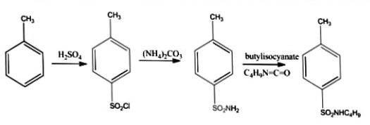
  • Synthesis of Tolbutamide

    Aim: To prepare and submit Tolbutamide from Toluene


    Reference: Jayaveera K.N., Subramanyam S., Reddy K.Y., Practical Medicinal Chemistry: S.Chand Publication; 2018, Vol. 53; Page no 397


    Chemicals Required: Concentratic Sulphuric acid, Toluene, Phosphorus pentachloride, Iodine Lime water, Sodium Carbonate, Ethyle chloroformate, Ammonium carbonate, Anhydrous potassium Carbonate, Acetic acid, dry acetone, Butyl amine, Butylisocynate


    Apparatus Required: RBE, Condenser, Beaker, Funnel, and Glass rod

    Principles: Tolbutamide, NP-methylbenezenesuphonyl)-N-butyl urea is an oral hyperglycemic
    agent used for the treatment of non-insulin-dependent diabetes mellitus (NIDDM). It is structurally similar to acetohexamide, chlorpropamide, and tolazamide and belongs to the sulphonylurea class
    of insulin secretagogues which act by stimulating the beta cells of the pancreas to release insulin.
    Sulphonylureas increase both basal insulin secretion and metal stimulated insulin release

    Synthesis of Tolbutamide
    Synthesis of Tolbutamide

    Procedure:

    Procedure for synthesis of p-toluenesulphonyl chloride from toluene

    • Heat 130 gm of pure toluene and 450 gm conc. sulphuric acid in a cast- iron pot fitted with suitable agitator. Add few crystals of iodine and allow the temperature to rise 100c.
    • The sulphonation is complete in about 6 hrs, transfer the reaction mixture in a large beaker diluted with water. Add lime water gradually neutralize the excess acid.
    • Filter out the calcium sulfate and any ferric hydroxide and wash with hot water.
    • Add sodium carbonate to the filtrate until just alkaline to phenolphthalein and remove the calcium carbonate by filtration. Evaporate the filtrate almost to dryness, when sodium salt of o toluene Sulphonyl and p-toluene sulphonyl acids separate out. When the reaction is complete add cold water. The p-toluene sulphonyl chloride separates as a solid and recrystallizes with alcohol. The O-toluene sulphonyl chloride is an oily liquid and is separated from the filtrate by means of a separating funnel.

    Procedure for synthesis of p-toluene sulphonamide

    • Thoroughly mix 5 grams of p-1oluene sulphonyl chloride and 10 grams of ammonium carbonate by grinding in a mortar until a fine powder is obtained.
    • Heat the mixture on a water bath with continuous stirring to convert sulphonyl chloride into sulphonamide. Crystallize the product from hot water. Filter off as colourless crystals.

    Procedure for the synthesis of ethyl p-toluene sulphonyl carbamate

    • Mix the corresponding sulphonamide 49 grams with ethyl chloroformate 42 grams and anhydrous potassium carbonate 3 grams in dry acetone 400 ml and reflux for 20 hours. Remove acetone by distillation under reduced pressure and keep the solution overnight and then add in water 250 ml. Neutralize with acetic acid. Filter the solid product and wash with distilled water and recrystallize with alcohol.

    Procedure for synthesis of tolbutamide

    • Dissolve the specified carbamate derivative in hot toluene 30 ml. Add butylamine slowly and reflux for 4 hours. Filter and dry it.

    Report: Tolbutamide was prepared and submitted.

    Uses: Tolbutamide is used together with diet and exercise to improve blood sugar control in adults
    with type 2 diabetes mellitus. Tolbutamide is not for treating type I diabetes.

  • Synthesis of Hexamine

    Aim: To prepare and submit Hexamine

    Chemicals Required: Distill Water, Formaldehyde, Conc: NH3 or NH4OH, Diethyl ether and ethyl alcohol.

    Apparatus Required: Rotary Vapouriser, Thermometer, Litmus Paper, Measuring Cylinder, Reflux Condenser, Round Bottom Flask, Beaker, Buchner Funnel, Water Bath, Vaccum Desiccator and Glass Rod.

    Principle: Hexamine is a heterocyclic organic compound that is also known as Methenamine or Hexamethylenetetramine or Urotropin. Hexamine use as an anti-infective agent and used to treat Urinary Tract Infections. Hexamethylenetetramine is prepared by the condensation of formaldehyde and ammonia

    Synthesis of Hexamine
    Synthesis of Hexamine

    Procedure: The procedure of preparing hexamine is given below:

    1. Place of 10ml of 37% formaldehyde and 9ml of ammonia into a round bottom flask.
    2. Evaporate the solution on a Rotavapor.
    3. During evaporation, a white residue (urotropin) is formed.
    4. Add another portion of Ammonia 14ml to dissolve the solid.
    5. Once more place the flask onto a Rotavapor and evaporate the solvent. Repeat the solvent. Repeat that step once more with an additional portion of ammonia(14ml).
    6. After evaporation add 15ml of anhydrous ethanol and dissolve the solid by heating under reflux.
    7. Filter the hot mixture on the Bichner Funnel.
    8. Add 10ml diethyl ethar to the filtrate and cool down.
    9. Filter the precipitated urotropin wash it with anhydrous ethanol drain well and then dry on air.

    Report: Hexamine was prepared and submitted.

    Uses of Hexamine: Uses of Hexamine is discussed below:

    • Hexamine dominantly used in the preparation of powdery and liquid preparation of phenolic resin moulding compounds.
    • Hexamine is also used as a food preservatives.
    • Hexamine is also use against concomitant odour and excessive sweating.