Author: Pankaj Saha

  • Study The Effect Of Hepatic Microsomal Enzyme Induction

    Aim: To study the effect of hepatic microsomal enzyme induction on the duration of action of
    pentobarbital sodium.

    Principle: The drugs induce hepatic microsomal oxidative enzyme systems to enhance the metabolism of other drugs. as a result, in the presence of an enzyme-inducer, the duration of action of the second drug will be reduced. This has significant clinical relevance because when more than one drug is administered at a time one drug may modify the action of another through the microsomal enzyme-inducing property.
    The common drugs which induce hepatic microsomal enzyme systems are phenobarbitone and meprobamate. Co-administration of any drug with either of these drugs may affect the disposition of the second drug and therefore, the desired pharmacological effects. The students are advised to study various processes of drug metabolism before doing this experiment.

    Requirement:
    Animal: Mice(20-25g)
    Drugs: Phenobarbitone sodium (dose: 50mg\kg IP, once a day for 5 days. Prepare a stock containing 5mg\ml of the drug and inject 1ml\100g of body weight of the animal. Prepare fresh solution every day). Pentobarbital sodium (dose45 mg\ml of the drug and inject 1 ml\100 g of body weight of the animal).

    Procedure:

    1. Weigh and number the animals. divide them into two groups, each comprising of at least 6
      mice.
    2. To the first group inject phenobarbitone once daily for 5 days. To the second group inject
      distilled water, similarly, for 5 days.
    3. One hour after the last dose of phenobarbitone on the 5th day, inject pentobarbital to both the groups.
    4. Note the onset and duration of sleepdue to pentobarbital in both the groups as done in the
      earlier experiment.

    The inference the animals pretreated with phenobarbital sleep for a shorter duration of the time as
    compared to animals treated with distilled water.

  • Anticonvulsant Effect Of Drugs By Maximal Electro-shock Induced Convulsions In Rats

    Aim: To study the anticonvulsant activity of phenytoin against maximal electro shock-induced
    convulsion rats.

    Principle: Different types of epilepsy i.e., grand mal, petit mal or psychomotor type, can be studied in
    laboratory animals. The maximal electroshock (MES) induced convulsions in animals represent
    grand mal epilepsy. Similarly chemo- convulsion due to pentylenetetrazole which produce the clonic
    type of convulsion in man. These are the two procedures used to study convulsion and to test anticonvulsant drugs in laboratory animals.
    In MES- convulsion electroshock is applied through the corneal electrodes. Through optic
    stimulation, cortical excitation is produced. The MES convulsion is divided into five phases are

    1. Tonic flexion
    2. Tonic extensor
    3. Clonic convulsion
    4. Stupor
    5. Recovery / death
      A substance is known to posses anti convulsant property if it reduces or abolishes the
      extensor phase of MES convulsions both in rats and mice. It is advised the students should
      have complete background of the pharmacology of anti epileptics drugs before performing
      this experiment.

    Requirement:
    Animal: Rats (150-200 gm)
    Drugs: phenytoin (dose 25 mg/kg); prepare a stock solution
    Equipment : electro- convulsiometer, cornel electrode (150 mA current for 0.2 sec) Stop watch

    Procedure:

    1. Weigh and number the animals. Divide them into two groups each consisting of 4-5 rats.
      One group is used as control and the other for drugs treatment.
    2. Hold the animals properly , place corneal electrodes on the cornea and apply the
      prescribed current. Note the difference stages of convulsion
    3. Repeat with other animals of control group
    4. Inject phenytoin I.P to group of 4-5 rats. Wait for 30 min and subjected to animals to
      electroconvulsions .
    5. Note the reduction in time or abolition of tonic extensor phase of MES convulsions.
  • Study Of Drugs On Locomotor Activity Using Actophotometer

    Aim: To study the central nervous system depressant property of chlorpromazine on the locomotor activity of mice using an actophotometer.

    Principle: Most of the central nervous system acting drugs influencing the locomotor activities in man and animals. The CNS depressant drugs such as barbiturates and alcohol reduce motor activity while the stimulants such as caffeine, amphetamines increase the activity. In other words, the locomotor activity can be an index of wakefulness of mental activity. The locomotor activity can be easily measured using an actophotometer. which operates on photoelectric cells which are connected in a circuit with a counter, when the beam of light falling on the photocell is cut off by the animal, a count is recorded. An actophotometer could have either a circular or square arena in which the animal moves. Both rats and mice may be used for testing in this equipment.

    Requirement:

    Animal: Mice (20-25gm)
    Drugs: Chlorpromazine hydrochloride
    Equipment : Actophotometer

    Procedure:

    • Weigh the animals and number them.
    • Turn on the equipment and place individually each mouse in the activity cage for 10 min. Note the basal activity score of all the animals.
    • Inject chlorpromazine and after 30 min re-test each mouse for activity scores for 10 min. Note the difference in the activity before and after chlorpromazine.
    • Calculate percent decrease in motor activity.
  • Effects Of Skeletal Muscle Relaxant Using Rotarod Apparatus

    Aim: To study the muscle relaxant property of diazepam in mice using rotarod apparatus.

    Principle: One of the important pharmacological actions of anti-anxiety agents of benzodiazepam class of drugs is muscle relaxing property. The skeletal muscle relaxation together with a taming or
    calming effect, these agents reduce anxiety and tension. The loss of muscle grip is an indication of
    muscle relaxation. This effect can be easily studied in animals using inclined planes or rotating
    rods.

    The difference in the fall off time from the rotating rod between the control and diazepam
    treated animal is taken as an index of muscle relaxation. The angle of the slope of the inclined
    plane or the rate of rotation of the rod should be adjusted such that a normal mouse can stay on
    the plane or on the rod for an appreciable period of time.

    Requirement:

    Animal : Mice (20-25gm)
    Drugs: Diazepam of the drug and inject 1ml/100g of body weight of the mouse.
    Equipment: Rotarod apparatus

    Procedure:

    • Weigh the animals and number them.
    • Turn on the instrument select an appropriate speed (20-25rpm)
    • Place the animals one by one on the rotating rod. One can place more than one mouse. Note down the fall-off time when the mouse falls from the rotating rod. A normal mouse generally falls off within 3-5 min
    • Inject diazepam to all animals. After 30 min repeat the experiments as done in step 3 note the fall off time.
    • Compare the fall off time of animals before and after diazepam treatment.
  • Effect Of Drugs On Ciliary Motility Of Frog Oesophagus

    Aim: To study the effect of physostigmine and atropine on ciliary movement in frog buccal
    cavity.

    Principle: Cilia in the buccal cavity and in the oesophagus helps in the movement of food particles. Similarly, the importance of mucociliary function has been established in the respiratory tract and of pulmonary diseases such as chronic bronchitis, asthma and cystic fibrosis. The integrity of mucociliary function is very important in these airway diseases. Cilia exhibit a great degree of autonomy in that they are capable of functioning in the absence of nervous innervations. It has also been demonstrated that the Ach present in the mucous membrane of the trachea and buccal cavity helps in the ciliary movement. Ach serves as a local hormone and the presence of choline acetylase support the fact that Ach is locally synthesised in the mucous membranes.

    Requirements:

    Animal: Frog
    Drugs: Physostigmine stock solution (1µg/ml), Atropine stock solution (1µg/ml)
    Physiological solution: Normal saline

    Procedure:

    • Decapitate the frog and pin the frog to the frog board on its back.
    • Pin the lower jaw to the abdomen cutting sufficiently the buccal cavity and exposing the oesophagus wet by irrigating it with normal saline.
    • To assess the distance travelled by the particles, fix two points. One starts from the lower jaw to the other beginning of the oesophagus. Keep this distance constant to measure the time taken by the particle to move from a fixed point in the lower jaw to the beginning of the oesophagus.
    • Place a poppy seed or a small piece of cork at the premarked spot in the jaw. Turn on the stop watch and note the time taken by the object to reach the beginning of the oesophagus repeat this several times.
    • Put a few drops of physostigmine on the buccal cavity and after 10 min repeat step 4 note the tome.
    • Wash the buccal with normal saline. Put a few drops o atropine on the buccal cavity. After 10 min repeat step 4. Note the time.
    • Find out the differences in the time taken by the object to move between the premarked distance in the buccal cavity in presence of saline, physostigmine and atropine.

    Inference: Physostigmine reduces and atropine enhances the time taken by the object to move
    from the pre-marked point in the lower jaw to reach the oesophagus, respectively.

  • Effect of Drugs on Rabbit Eye

    Aim: To study the Mydriatic effect of topically applied atropine on rabbit eyes.

    Principle: A large number of drugs are used for their local action in the eye as eye drops or eye ointments. Most of these drugs belong to an antimicrobial, autonomic or local anaesthetic group. The eye is supplied by both sympathetic and parasympathetic nerves. The superior palpebral muscle and the dilator papillae of the iris have sympathetic supply. The sphincter pupillae of the iris has a parasympathetic supply that exercises dominant control. The ciliary muscle is also supplied by the parasympathetic nerve and when it contracts, the ciliary body is moved inwards and forwards. Because the lens bulges forward and the eye is accommodated for near vision. The opposite effect is produced by the relaxation of ciliary muscle resulting in paralysis of accommodation. When the pupil dilates, the iris folds back near the opening of the canal of schlemn and the drainage of aqueous humour is decreased thereby increasing intraocular pressure. constriction of the pupil, by opposite action, will increase drainage and reduce intraocular pressure.

    Topically applied drugs can affect the eye by changing conjunctival congestion, papillary size, light reflex, corneal sensitivity and intraocular pressure. however, the effects of drugs on papillary size, light reflex and corneal reflex can be easily studied by the students. the pupillary size can be measured by placing a transparent plastic scale in front of the eye but as close as possible. Light reflex is elicited by directing the light of a torch towards the pupil. The sensitivity of the cornea is tested by gently touching the cornea with a fine cotton swab stick from the side and not from the front of the eye. This elicits corneal reflex which manifests as the blinking of the crystals.

    Requirements:

    Animal : Rabbit(2-5kg)

    Drugs: Atropine (1%w/v) & Physostigmine (1%w/v)

    Equipment: rabbit holder, pen torch

    Procedure:

    • Place the rabbit in the rabbit holder by keeping the head outside.
    • Observe size of the pupil in both the eyes.
    • Examine the effect of light reflex by holding the torch in front of the eye moving light beam to and fro
    • Examine the corneal reflex by touching a side of the cornea with a cotton swab.
    • Instil a few drops of atropine solution in the conjunctiva over a period of 8-10 min in the right eye of the rabbit would serve as the control. Instil normal saline in the left eye.
    • Record the pupillary size, light reflex and corneal reflex after ten minutes of drug initialisation and tabulate the observation.
    • Repeat the experiments with physostigmine and ephedrine.

    Observation:

    DRUGPUPILLARY
    SIZE
    LIGHT REFLEXCORNEAL
    REFLEX
    SalineNormalPresentPresent
    PhysostigmineConstrictionPresentPresent
    EphedrineDilationPresentPresent
    AtropineDilationPresentPresent
  • Study of Different Route of Drug Administration

    There are different routes of drug administration by using laboratory animals.

    Aim:

    • To learn how to handle, treat preparing animals for experiments and ethical guidelines during the treatment.
    • Measure the required volume of drugs using aspectic techniques.
    • To learn how to give a different types of route of drug administration.

    How to handle: Good handling and restraint are the most important techniques for correct administration. The restrain methods are 2 types.

    • Physical method
    • Manual method
      • Double handled manual method
      • Single handled manual method

    Route of drug administration:

    • Enternal route (oral and intregastric)
    • Parentral (I.V, I.M,I.P,S.C,I.D)
    • Topical route (surface of the skin membrane)

    Enternal route: The placement of drugs directly into any part of GIT directly it could be oral, sublingual, intragastric.

    Oral route: The swallowing of a drug through the mouth by the desired drug along with water (ad libitum) or food. It is economic and convenient, safe and some animals can be trained to cooperate
    voluntarily. This route is not referable since it is inaccurate.

    Intragastric route: The administration of fluid/drugs directly into the oesophagus or stomach is often used instead of mixing the substance in water or food. This method is accurate for the dosing od
    animals.

    The gastric lavage tube is a small curved, metallic tube with the ball on the tip, it is often used
    without anaesthesia.

    Parental route: Subcutaneous route: the part of the subcutaneous injection and back of the neck it doesn’t require a large volume of drugs suitable for suspension.

    Intraperitoneal route: Commonly used in rats and mice, rapid absorption due to the large particle surface, the mouse may be injected compared with rat (ranges from 2-10ml/kg ).

    Intravenous route: It is the most efficient route of drug administration. It should be used with the help of restraining devices in the appropriate size for the animals to be injected. The tail vein is suitable
    for drug administration.

    Topical route: The most convenient route for topical drugs, drugs like local anaesthetics, ointments and cream can be easily applied on the surface of the skin membrane. Eye drops and ointments were also used in this method. Animals like rabbits, guinea pigs and rats are mainly used in this method.

  • Study of Common Laboratory Animals

    In this post, we will learn about the different types of common laboratory animals which we often use in our Pharmacology Lab.

    Guinea pig

    Guinea pigs (400-600g) are the commonly used experimental animals. they are very docile and easy to raise and maintain they are highly sensitive to histamine. They are used in experimental asthma to study bronchodilators. they are also used to local anaesthetics and as a model in amoebiasis and cholera as they are sensitive to this microorganism.

    Albino rat

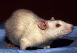
    The white rat (200-250g) is the commonest laboratory animal used in experimental pharmacology. Rats are easy to breed and maintain. Resemble man in several organ function and nutrition and sensitive to most of the drugs; make them very useful experimental animals. However, they do not have a vomiting centre. The various rat tissue used is colon, stomach, uterus, caecum and vas deference. Besides these organs rat brain tissue is extensively employed in radio receptor-ligand studies. The other strains of rats are Sprague- Dawley and porton.

    Albino mouse

    White mice are the smallest laboratory animals used. Mice are also easy to breed and maintain. They are small in size (25-30g) and therefore, easy to breed and maintain. They are sensitive to most of the drugs used in experimental pharmacology. Mice are used extensively in toxicity study, bioassay of insulin, testing of analgesics, CNS active drugs and chemotherapeutic agents. More recently mouse brains, as well as primary cell culture ofmouse spinal cord neurons, are used in neuropharmacology for studying neurotransmitters receptor functions the other strains of mice used are laca and balb/C.

    Rabbit

    Domestic rabbits (2-3 kg) are generally used for pyrogen testing. Some of the tissues or organs from rabbits used are the heart, aorta, duodenum and ileum. One peculiar thing about rabbits is that they are resistant to the actions of atropine as they contain the atropine-esterase enzyme, the presence of which is genetically determined.

    Frog

    Frogs (150-200g) were one time extensively used in experimental pharmacology. However, recently the use of wild frogs for experimental purposes has been banned. Earlier, frogs were used for isolated heart, rectus abdominis muscle preparation, the study of muscle nerve and ciliary movements, respectively. Frogs were also used for the study of nerve block type of local anaesthetics. frogs are inexpensive and easily available, and the ban on the use of frogs has been debated.

    Other animals

    Cats, dogs and monkeys are used for pharmacological investigations of drugs. Cats and dogs were one time commonly used to study blood pressure experiments. But their use has been now restricted. However, beagle dogs are the only strain approved by regulatory authorities (USFDA) for preclinical testing of new drugs.

    Alternatives to animal experimentation

    Because of the growing concern about the use of animals in biomedical research in several countries have passed legislation to prevent or usage of animal experimentation. these include experiments with tissue and body fluids of normal animals and human use of microorganisms, primary cell culture and cell lines, use of models and computer simulation and software are being used nowadays. As per the common laboratory alternate animals.

    Common NameScientific NameUsed In
    Zebra fishDanio rerioDevelopmental assay, Drug discovery, Neurobiology, Toxicity
    WormsHabolitisDevelopmental assay, Neurobiology, Toxicity
    Fruit flyDrosophilaCancer biology, Developmental assay, Neuroscience
    TunicatesCiona spDevelopmental assay, Neuroscience
    StarfishEchinodermsDevelopmental assay, neuroscience

  • Commonly Used Instruments In Experimental Pharmacology

    In spite of tremendous development in electronic devices and recording systems are followed in institutions and research laboratories. Some common instruments are used in experimental pharmacology.

    Organ bath:

    Source: Wikipedia

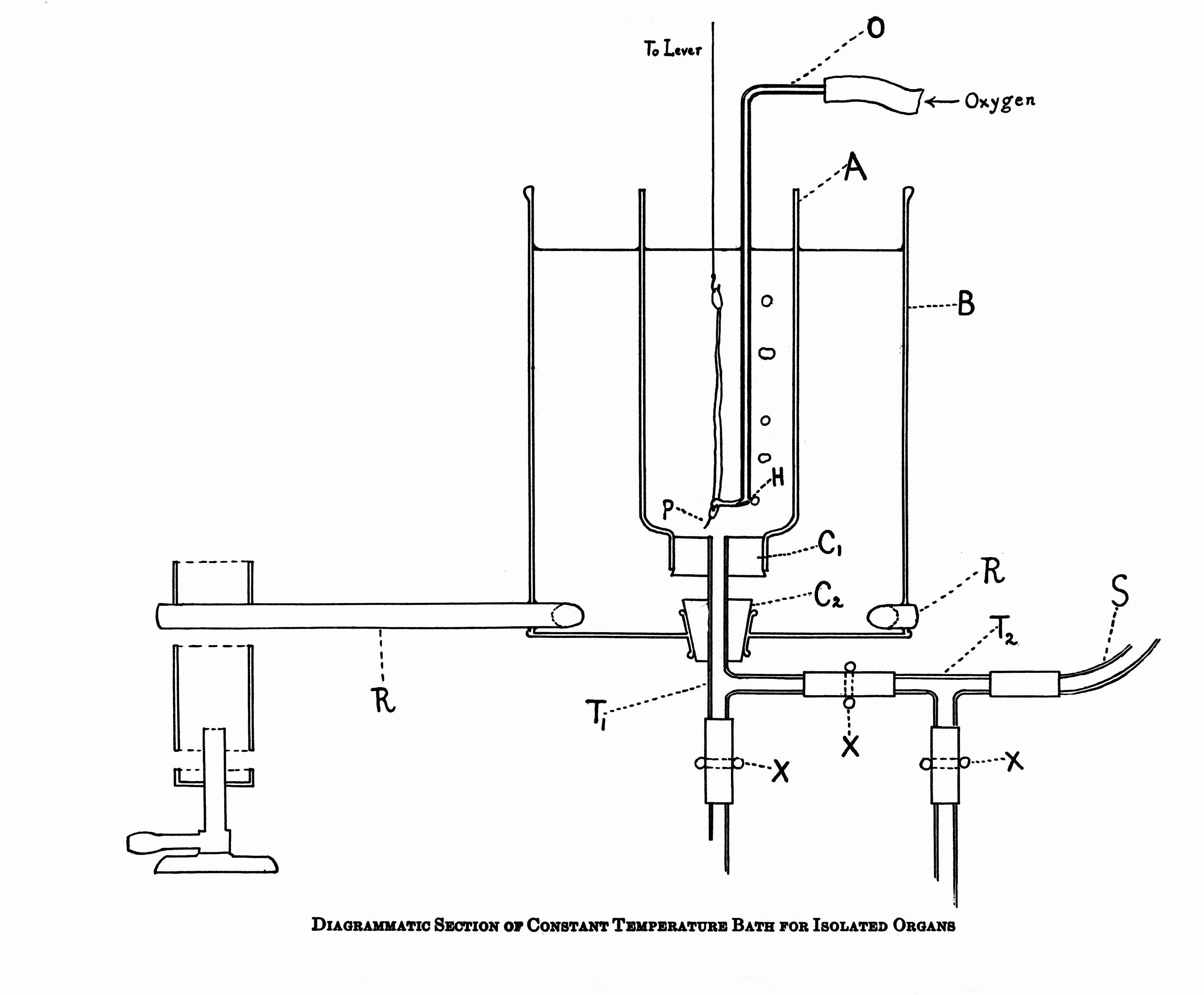
    The tissue bath used to put the animal tissue for studying the drug actions is called student organ bath. This was first designed by Rudolph Magnus in 1904. The organ bath essentially
    consists of

    • An outer jacket made of up of steel or glass or Perspex.
    • The inner organ or tissue bath made up of glass with a capacity varying from 10 -50 ml.
    • Thermostatically controlled heating rod.
    • Stirrer to keep the water in the outer jacket at uniform temperature.
    • Oxygen or delivery glass tube which also serves as tissue holder.
    • Glass coil, one end of which is connected having the physiological salt solution. The student organ bath having two units of inner tissue bath is called double unit organ bath.

    Rotarod apparatus:

    Source: Wikipedia

    For the study of muscle relaxant property of diazepam in mice. The loss of muscle grab is an indicator of muscle relaxation. This effect can be studied in animals using an inclined plane or rotating rods.

    Actophotometer:

    Source: ResearchGate

    It is used to study the CNS depressant property of chlorpromazine on the locomotor activity of mice. CNS depressant drugs like alcohol reduce motor activity with the stimulants like caffeine and amphetamines increase the activity. The actophotometer operates on the photoelectric
    cell which is connected to the circuit with a counter.

    Electro-convulsiometer:

    It is used to study the anticonvulsant activity of phenytoin against electro-convusiometer induced in rats. The electric shock is applied through the corneal electrodes; it produces 5 phases such as tonic flexion, tonic extensor, clonic convulsion, stupor, and recovery/death.

    Pole climbing apparatus:

    It is used to study anxiolytic activity in rats and mice. The basic principles of pole climbing apparatus are based on a neurochemical mechanism of anxiety disorders drugs like benzodiazepam are used.

    Analgesiometer:

    Source: Solution Pharmacy YouTube Channel

    It is used to study the analgesic effects of drugs in mice and rats. The inducer of pain such as heat, physio compression and chemical inducers. The basal reaction time was noted as an inference of pain sensation drugs like morphine and other NSAIDs are used.

    Metabolic cage:

    It is used to study the metabolic parameters such as faecal and urine for the study of
    purgative or laxative, animals such as rats and mice are used.

  • RBC Count

    Aim: To find out the number of red blood cells in one cubic millimetre of blood.

    Principles: The number of RBC in a known volume of diluted blood is counted and the number of
    cells in one cm of undiluted blood are calculated from this.

    Apparatus Required: Hemocytometer, RBC diluting fluid, compound microscope, sterile lancet,
    watch glass, cotton, rectified spirit.

    Uses of R.B.C. pipette:

    • WBC count in leukemias
    • Platelet counting

    Uses of the bead in the bulb:

    • For proper mixing
    • To know whether the pipette is dry
    • To identify the pipette
    • R.B.C diluting fluid: Hayem’s fluid is the commonly used diluting fluid.

    Composition:

    • Sodium chloride 0.5 Gm
    • Sodium sulphate 2.5 Gm
    • Mercuric perchloride 0.25 Gm
    • Distilled water 100 ml sodium chloride and sodium sulphate together keep the isotonicity of fluid. Sodium sulphate also prevents the clumping of red cells. Mercuric perchloride fixes the cells and acts as a preservative.
    • Other diluting fluids:
      • Gower’s fluid
      • Toison’s fluid
      • Formol Citrate solution

    Procedure: Clean and dry the counting chamber and put on the special coverslip provided. Focus on the high power objective and identify the RBC counting area. Clean the RBC pipette first with distilled water, then with absolute alcohol and finally with ether and keep it dry. Take a small quantity of diluting fluid in a watch glass and keep it aside. Clean the fingertip using rectified spirit and make a deep prick with a sterile lancet, so that blood comes out freely without squeezing. Wipe off the first drop which may contain tissue fluid also. Allow a good-sized blood drop to form a hanging drop and
    keep the pointed tip of the pipette touching the drop. Suck in blood up to the 0.5 marks carefully, without any air bubble. Excess blood at the tip of the pipette is removed using a blotting paper or piece of cotton. Immediately, diluting fluid from the watch glass is sucked in up to the 101 marks without any air bubble by keeping the pipette in a vertical position.

    Then thoroughly mix the blood and diluting fluid in the pipette by gently rolling the pipette held horizontally between the palms and keep aside. Mixing takes place only in the bulb of the pipette. The column of diluting fluid contained in the stem of the pipette does not enter into the dilution (i.e. 101-1 = 100). So that the blood sucked up to the 0.5 marks will have a dilution of 0.5 in 100 or 1 in 200. Now take out the counting chamber for charging discard the first few drops from the pipette, as the stem contains only diluting fluid. Bring one small drop of diluted blood at the tip of the pipette, to the edge of the coverslip on the counting chamber at an angle of about 450 The fluid enters by capillary action under the coverslip and fills the counting chamber. Both areas are filled.

    Focus the RBC counting area under high power. Keep the counting chamber undisturbed for about 3 minutes for the cells to settle down in the counting area, and start counting. At least 5 squares, each having 16 smallest squares (preferably 4 corners and 1 central) should be counted to obtain a satisfactory average and a better dispersal value. While counting each small square, cells touching the top and left margin of each square should be omitted and cells touching the bottom and right margin of each square should be counted. Draw a chart of the counting squares in the record and enter the number of cells in each square and when counted.

    Calculations: Let the number of cells counted in (5×16) 80 smallest squares be “N”

    The number of cells in 1 smallest square is N/80

    Side of 1 square = 1/20mm

    Side of 1 square = 1/20mm

    The volume of diluted blood in 1 square=1/400×1/10=1/4000mm3

    Number of cells in 1/4000mm3 diluted blood = N

    80 Number of cells in 1 mm3 of diluted blood N 80×1/4000= Nx4000 80

    The dilution factor is 1 in 200

    (Total diluted volume in the bulb of the pipette is 100 parts, out of which 0.5 is blood. So dilution is 0.5 in 100 i.e.1 in 200)

    So numberof cells in 1 mm3 of undiluted blood =Nx 4000×200 = Nxl0000 80

    Precautions:

    • Counting chamber and pipette should be clean and dry.
    • Fingertip and pricking lancet should be sterile.
    • Blood should freely come out without squeezing.
    • Be careful to prevent clotting of blood inside the pipette.
    • While filling the pipette and charging the counting chamber, no air bubble should enter.
    • Blood should be taken only up to the 0.5 marks and diluting fluid sucked only up to 101 mark.
    • Blood should be properly mixed with the diluting fluid.
    • Discard first few drops before charging because it will not contain RBC5.
    • While charging the counting chamber, over filling and spilling should be avoided.
    • Cells should be settled down and more or less evenly distributed before counting.
    • Don’t keep the microscope in tilted position.
    • Count from Left to Right and avoid counting of the same cell.